It is known that almost every person is faced with domestic or sports injuries, which result in damage to the ligaments of the joints. Ligaments are fibers of dense connective tissue, the damage of which during life is almost not restored on its own. A socially significant disease is rupture of the anterior cruciate ligament of the knee joint. According to Russian medical institutions, the incidence of injuries to the anterior cruciate ligament is one in 200 people, with the majority of patients being active and able-bodied people aged 16 to 45 years. Against the background of damage to the ligaments, concomitant diseases develop, such as joint instability, synovitis, gonarthrosis, which can lead to disability. For the treatment of ligament rupture, it is necessary to perform arthroscopic knee surgery, i.e. carry out a complete replacement of the torn ligament. To do this, it is necessary to form bone canals in the femur and tibia and connect them with a graft or ligament implant. Autografts (tissue fragments of the patient himself), allografts (tissue fragments of donors, including animals), as well as artificial implants can be used. The use of autografts is the most effective solution, however, to obtain them, tissue must be removed from another part of the body, causing additional trauma. When transplanting allografts, there is a high probability of rejection or transmission of infections to the patient from the donor. Thus, the future of replacing damaged ligaments with synthetic materials. For the manufacture of artificial ligaments, polymer threads are used, which are braided in a certain way into fibers. One of the important reasons for the unsatisfactory results of surgical treatment of ligament injuries is the poor biological integration of the canal wall with such implants. This is due to the fact that the mechanical characteristics of the fibers of the implant exceed the strength of the bone tissue of the channel in which it is fixed. In this regard, there is a problem of destruction of the bone canal, especially in the first time after the operation. In addition, the polymers used for the manufacture of ligaments are hydrophobic and chemically inert materials with low biological properties, which are difficult to take root in the bone canal.
To overcome the above problems, Russian scientists have developed a new composition and method for forming a coating for polymer fibers of a binder. Representatives of the Saratov University, the National Research University "Moscow Institute of Electronic Technology" (MIET), the First Moscow State Medical University named after I.M.Sechenov (Sechenov University), the Institute of Microelectronics Nanotechnologies of the Russian Academy of Sciences, the Moscow Regional Research Clinical Institute named after MF Vladimirsky, Russian State Agrarian University - Moscow Agricultural Academy named after K.A. Timiryazev, Federal State Budgetary Scientific Institution "Scientific and Production Complex" Technological Center ". The resulting coating is a carbon nanotube framework structure formed by infrared radiation in a collagen matrix. Before starting to manufacture the coating, extensive theoretical studies were carried out, which made it possible to select nanotubes and protein molecules with the necessary parameters that would provide an energetically efficient composition of the coating. These studies were carried out by the research team of Professor O.E. Glukhova using molecular dynamics and the quantum mechanical density functional method in the tight binding approximation with a self-consistent charge SCC DFTB. The results of computer studies made it possible to establish the optimal temperature and time for the formation of equilibrium configurations of complexes based on single-wall carbon nanotubes and collagen. The selected nanotubes have a diameter of about 1.5 nanometers and allow the formation of a high-resolution framework structure. Due to the functional chemical groups of nanotubes, a high hydrophilicity of the coating was achieved, which significantly exceeds the hydrophilicity of polymer fibers and a coating of pure collagen. Due to this, synthetic bonds with nanocoating ensured a normal level of blood hemolysis. Those. upon contact of the nanocoating with blood, the destruction of the membranes of erythrocytes above the natural level does not occur. Studies have shown that the coating is biodegradable when exposed to body fluids. At the same time, structural changes occur in it, namely, the number of pores and their size increase. This is necessary for the growth of cells, blood vessels and nerve fibers into the structure of the coating to ensure better integration of the implant with the surrounding tissues.
The nanocoated bundles were tested on laboratory animals. The success of joint ligament reconstruction depends on the effectiveness of the healing of the bone canal and fixation of the artificial ligament in it within a few months after implantation. Scientists have shown that the developed ligaments with nanocoating ensure the fastest growth of bone and dense connective tissue in the canal during the first 6 months after surgery. Due to this, it was possible to achieve tight fixation and complete engraftment of the synthetic ligament with nanocoating in comparison with the implant made of traditional polymer fibers, which are usually used by orthopedists. As a result of the research, an important mechanism of interaction of the coating with biological tissues was determined. The peculiarity of the mechanism was the appearance of a liquid medium around the synthetic ligament with nanocoating, in which the loosening reticular tissue initially germinated. Then it was replaced by dense collagen fibers, which in turn were replaced by bone. The importance of reliable integration of the ligament into the bone canal is due to the fact that the natural connection of the ligament to the bone is a highly specialized and organized structure that transfers mechanical stress from soft tissues to bone.
Thus, the use of the developed nanocoated joint ligament implant will allow achieving complete and prompt postoperative recovery of the patient.
The results of the joint work of SSU scientists with a number of leading scientific organizations of the Russian Federation have been published in the top-rated International Journal of Molecular Sciences published by MDPI. Immediately after its appearance on the magazine's website, the article Frame Coating of Single-Walled Carbon Nanotubes in Collagen on PET Fibers for Artificial Joint Ligaments aroused keen interest among the magazine's readership. In the first few days of its stay, the article has already collected 250 views and more than 100 downloads.
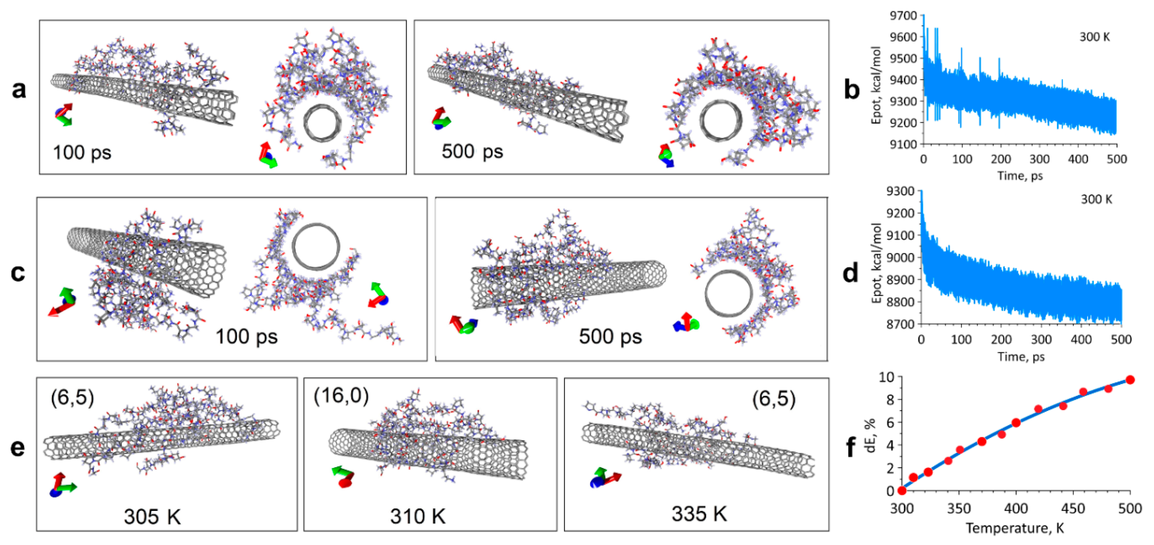
Figure: 1. Results of a molecular dynamics study of the formation of an equilibrium configuration of a single-walled carbon nanotube / collagen complex.
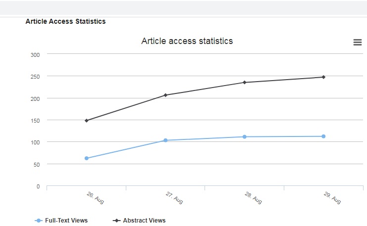
Figure: 2. Statistics of views and downloads of the article Frame Coating of Single-Walled Carbon Nanotubes in Collagen on PET Fibers for Artificial Joint Ligaments, taken from the official website of the International Journal of Molecular Sciences published by MDPI.